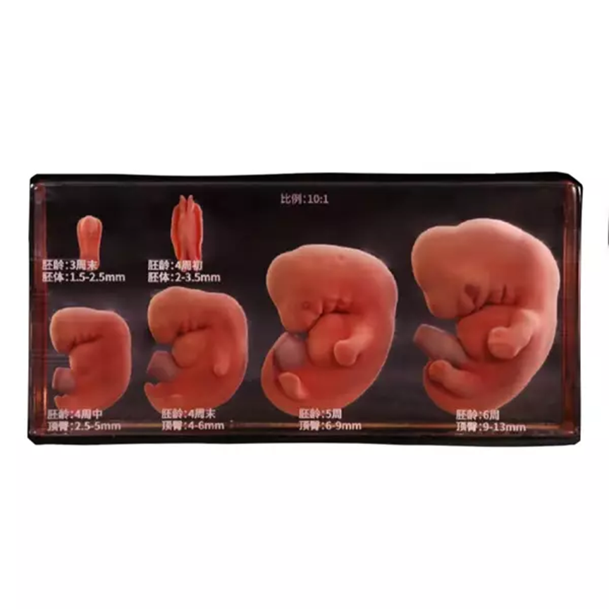
Assuredly, visualizing the intricate processes of embryological development can be challenging. DIGIHUMAN addresses this challenge with its advanced embryo 3D model, providing a detailed and interactive representation of early human development. This innovative tool is designed to enhance medical understanding and improve training for physicians, medical students, and researchers.
Comprehensive Anatomical Structures
DIGIHUMAN’s embryo 3D model incorporates a comprehensive range of anatomical structures, including bone, muscle, blood vessels, nerves, and ligaments. The model is derived from high-precision digital human data, ensuring accuracy and realism. The use of original sectional data, refined segmentation data, and three-dimensional geometric models allows for a detailed exploration of the developing embryo.
Model Making Process
The model-making process involves extracting voxels from the volume data on the surface of each anatomical structure. This ensures that the geometric model accurately reflects the appearance of real anatomical specimens. The texture map of the model is carefully generated to provide a visual perception consistent with cadaveric specimens fixed with formalin. This meticulous attention to detail makes DIGIHUMAN’s embryo 3D model a valuable tool for medical education and research.
Conclusion
In essence, DIGIHUMAN’s embryo 3D model offers a significant advancement in medical education by providing a detailed, accurate, and visually consistent representation of embryonic development. By combining high-precision data with advanced modeling techniques, DIGIHUMAN is enhancing medical understanding and empowering the next generation of medical professionals.